CALEC surgery, short for cultivated autologous limbal epithelial cells, is paving the way for innovative approaches in eye damage treatment. Developed at Mass Eye and Ear, this groundbreaking procedure utilizes stem cell therapy to restore the cornea’s surface after severe injuries that traditionally rendered standard corneal transplants ineffective. By harvesting limbal epithelial cells from a healthy eye, CALEC surgery allows for the creation of a personalized cellular graft that can be expertly transplanted into the damaged area. This method not only provides hope for patients suffering from corneal trauma but also represents a significant advancement in ocular transplantation techniques. The clinical trials have demonstrated an impressive success rate, showcasing CALEC as a promising alternative for those with previously untreatable conditions caused by corneal damage.
Cultivated autologous limbal epithelial cell (CALEC) procedures embody a revolutionary method in ocular repair, focusing on the restoration of the corneal surface through stem cell applications. This innovative treatment extracts healthy stem cells from one eye and cultivates them to create a graft, effectively addressing conditions that compromise the cornea’s integrity. As an alternative to traditional corneal transplants, CALEC surgery exemplifies the potential of personalized medicine in treating ocular damage caused by traumatic injuries or disease. The use of limbal epithelial cells not only aids in repair but enhances the possibilities for better visual outcomes in affected individuals. By employing cutting-edge solutions for corneal repair, the future of eye health is bright, offering new avenues for restoration that were previously deemed impossible.
Understanding CALEC Surgery: A Breakthrough in Eye Treatment
CALEC surgery, or cultivated autologous limbal epithelial cell surgery, represents a significant advancement in the treatment of corneal injuries that were previously thought to be untreatable. For patients suffering from limbal stem cell deficiency caused by trauma or chemical burns, CALEC offers a novel solution by transplanting stem cells from a healthy eye into the damaged one. This innovative technique not only shows promising results in clinical trials but also highlights the potential of stem cell therapy in ocular repair, providing hope for improved vision and quality of life for individuals with severe corneal damage.
In a recent clinical trial conducted by Mass Eye and Ear, CALEC surgery demonstrated over 90 percent efficacy in restoring the corneal surface among participants. The procedure involves harvesting limbal epithelial cells from a healthy eye, expanding these cells into a graft, and then transplanting them into the affected eye. This method utilizes advanced manufacturing processes developed through rigorous research, showcasing the vital role of collaboration between various institutions in advancing ocular transplantation methods.
The Role of Stem Cell Therapy in Corneal Repair
Stem cell therapy stands at the forefront of medical advancements, particularly in the field of ocular restoration. By utilizing limbal epithelial cells derived from a patient’s healthy eye, researchers can bypass the limitations associated with traditional corneal transplants, such as the need for donor corneas and the risks of rejection. This regenerative approach not only aims to heal the external surface of the eye but also addresses underlying issues associated with vision loss.
The success rate observed in the CALEC studies is a testament to how stem cell therapy can revolutionize treatments for corneal injuries. With a restoration success rate soaring to nearly 93 percent over the study’s timeline, it is evident that this innovative approach offers a feasible alternative to conventional methods of eye damage treatment. As research in this area progresses, the integration of stem cell therapies into clinical practice may significantly impact the future of ocular health.
Limbal Epithelial Cells: The Key to Eye Recovery
Limbal epithelial cells play a crucial role in the health and integrity of the cornea. These specialized cells, located at the eye’s limbus, are responsible for maintaining the corneal surface, promoting healing after injuries. When injuries deplete these cells, they cannot regenerate, leading to conditions like limbal stem cell deficiency, which severely compromises vision. Understanding the role of these cells underscores the importance of therapies like CALEC, which aims to replenish the corneal surface effectively.
The depletion of limbal epithelial cells can result from various factors, including trauma, chemical exposure, and infections. The CALEC procedure’s success in restoring these cells and repairing the corneal surface provides new avenues for patients previously resigned to a life of poor vision and discomfort. By harnessing the regenerative potential of limbal epithelial cells, eye care practitioners can offer a more targeted approach to treating severe eye conditions.
Ocular Transplantation: Future Implications of CALEC Surgery
Ocular transplantation has long been a critical facet of treating severe eye injuries, yet challenges such as donor availability and rejection rates complicate its implementation. CALEC surgery paves the way for a new dawn in ocular transplantation, addressing some of these limitations while enhancing patient outcomes. This innovative procedure signifies not just a treatment for corneal injuries but a paradigm shift towards regenerative medicine in ophthalmology.
The implications of CALEC surgery extend beyond mere restoration of the corneal surface; they encompass a broader vision for ocular care. Future studies aim to refine the process and explore allogeneic options—which would utilize limbal stem cells from cadaveric donors—potentially increasing accessibility to those in need. As research continues, CALEC surgery could redefine expectations for successful ocular transplantation, ultimately leading to more inclusive treatment options for various levels of eye damage.
Clinical Results: Promising Success Rates in CALEC Trials
The clinical trials surrounding CALEC surgery have yielded promising results, solidifying its place in the future of eye damage treatment. With over 90% of participants experiencing significant corneal surface restoration, the findings underscore the therapy’s efficacy and safety. These trials not only mark the ability of CALEC to improve vision but also highlight the positive patient outcomes that are critical to advancing ocular transplantation.
Moreover, follow-up assessments revealed that almost half of the participants achieved complete restoration of their corneas within three months, showcasing the rapid effectiveness of CALEC. As additional studies are anticipated to include larger patient populations and extended follow-up periods, the continued success of CALEC may inspire broader acceptance and adoption of stem cell therapies across the medical community.
The Future of CALEC Surgery and Its Impact on Eyecare
Looking ahead, the future of CALEC surgery appears bright as researchers continue to explore its viability and expand access for patients with eye damage. The ultimate goal is to gain FDA approval for the procedure, thereby enabling it to enter common clinical practice. This breakthrough could significantly alleviate the burden faced by patients suffering from severe cornea injuries and enhance the overall quality of ophthalmic care.
To achieve this goal, ongoing studies will need to focus on refining the graft manufacturing processes and exploring the potential for using donor-derived limbal stem cells. As trials aim to encompass a more diverse range of patients, the promise of CALEC surgery will undoubtedly shape the landscape of ocular treatments, providing new hope for those afflicted by previously untreatable corneal conditions.
Safety Profile of CALEC Surgery: A Comprehensive Analysis
The safety profile of CALEC surgery poses an important aspect of its evaluation in clinical settings. Preliminary results from the ongoing trials indicate a high safety standard, with no significant adverse events reported among participants. This reassuring data adds to the therapy’s credibility and supports the case for its future application in clinical ophthalmology.
Despite the apparent success, complications such as minor infections have been noted, illustrating the need for ongoing monitoring and careful patient management post-surgery. Understanding the safety and potential risks involved in CALEC procedures will be vital for practitioners as they consider its integration into routine eye damage treatment, ensuring patient welfare remains the top priority.
The Collaboration Behind CALEC: A Multi-Institutional Effort
The development and success of CALEC surgery are attributed to a collaborative effort among numerous prestigious research institutions. From Mass Eye and Ear to Dana-Farber Cancer Institute, these partnerships exemplify the power of interdisciplinary synergy in advancing medical innovations, particularly in specialized fields like ophthalmology. This cooperation has enabled researchers to hone the manufacturing processes and clinical trial protocols necessary for effective treatment applications.
Such collaborations are crucial for facilitating knowledge exchange and innovation in complex fields such as stem cell therapy and ocular reconstruction. The concerted focus on joint research efforts not only advances CALEC surgery but also contributes extensively to the broader realm of regenerative medicine, ultimately benefiting countless patients facing severe ocular conditions.
Patient Perspectives: Experiences with CALEC Surgery
The experiences of individuals who have undergone CALEC surgery provide valuable insight into its impact on their lives. Many patients report a newfound sense of hope and improved visual acuity following treatment, underscoring the psychological and emotional benefits that accompany regenerative therapies. Narratives from participants highlight the relief from persistent pain and visual disturbances, showcasing how innovative eye damage treatments can transform lives.
Moreover, patient testimonials regarding the post-surgery recovery process emphasize the importance of support systems in managing expectations and experiences. Acknowledging the variability in outcomes, these perspectives offer crucial feedback that can help refine future clinical practices and inform patient education regarding the potential of stem cell therapies in ocular care.
Frequently Asked Questions
What is CALEC surgery and how does it relate to stem cell therapy?
CALEC surgery, or cultivated autologous limbal epithelial cell surgery, utilizes stem cell therapy to repair damaged corneal surfaces. This innovative procedure involves taking healthy limbal epithelial cells from a patient’s unaffected eye, expanding these cells into a graft, and then transplanting the graft into the damaged eye to restore vision and relieve pain.
How effective is CALEC surgery in repairing corneal damage?
CALEC surgery has shown a remarkable effectiveness rate of over 90% in restoring the corneal surface in patients. In clinical trials, 50% of participants had their cornea completely restored by the three-month marker, with success rates increasing to 79% and 77% at 12 and 18 months, respectively.
What are limbal epithelial cells and their role in CALEC surgery?
Limbal epithelial cells are essential for maintaining the cornea’s smooth surface. In CALEC surgery, these cells are harvested from a healthy eye, expanded into a cellular graft, and transplanted into the damaged eye to replace lost or depleted cells, facilitating the regeneration of the corneal surface.
How does CALEC surgery address ocular transplantation challenges?
CALEC surgery provides an alternative to traditional ocular transplantation methods for patients with limbal stem cell deficiency. By utilizing a patient’s own stem cells, CALEC surgery increases the likelihood of graft acceptance and avoids complications associated with donor tissues, thereby enhancing the potential for successful restoration of vision.
Is CALEC surgery currently available for patients with eye damage?
Currently, CALEC surgery remains experimental and is not widely available at hospitals, including Mass Eye and Ear. Ongoing studies are necessary to confirm its effectiveness and safety before it can be considered for broad clinical use and submission for federal approval.
What are the potential risks associated with CALEC surgery?
While CALEC surgery has demonstrated a high safety profile, potential risks include minor adverse events such as infections. One case of bacterial infection was noted in a participant, highlighting the importance of post-surgery care, particularly regarding contact lens use.
What does the future hold for CALEC surgery and its accessibility?
Future plans for CALEC surgery include developing an allogeneic manufacturing process using cadaveric limbal stem cells. This advancement could potentially broaden the treatment’s availability, allowing options for patients with bilateral eye damage and enhancing overall access to this innovative therapy.
What is the significance of the CALEC surgery clinical trial?
The CALEC surgery clinical trial is significant as it is the first human study of stem cell therapy for ocular repair funded by the National Eye Institute. It represents a pioneering breakthrough in the treatment of corneal injuries previously deemed untreatable, potentially transforming the standard of care for patients with severe corneal damage.
| Key Points |
|---|
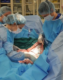
| Ula Jurkunas performs the first CALEC surgery at Mass Eye and Ear. |
| Stem cell therapy restores corneal surfaces in patients with eye injuries. |
| CALEC involves removing stem cells from a healthy eye and transplanting them into a damaged eye. |
| The method has shown a 90% success rate in restoring corneal surfaces. |
| Initial trials showed the procedure is safe with minimal adverse effects. |
| Future studies plan to include more patients and longer follow-up periods. |
Summary
CALEC surgery represents a groundbreaking advancement in the treatment of corneal injuries that were previously deemed untreatable. This innovative stem cell therapy has shown considerable promise in restoring corneal surfaces, leading to improved vision and quality of life for patients. As researchers continue to refine and expand this technique, there is hope for wider application, helping even more individuals regain their sight through the power of regenerative medicine.

